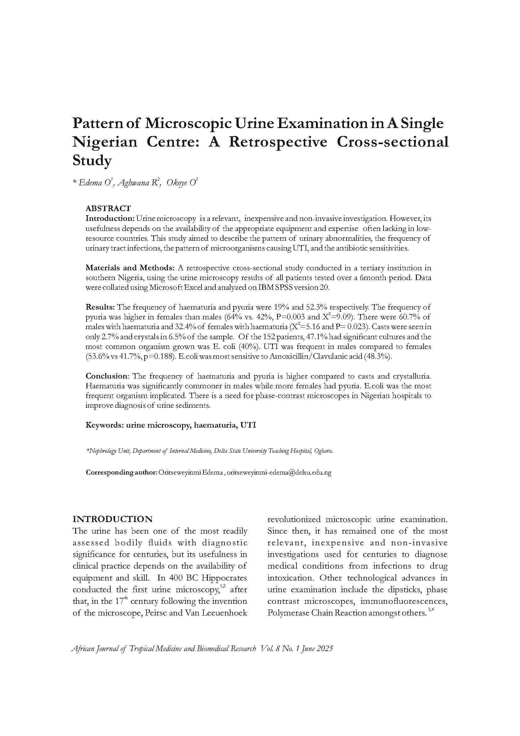
Pattern of Microscopic Urine Examination in a Single Nigerian Centre
A Retrospective Cross-sectional Study
DOI:
https://doi.org/10.4314/ajtmbr.v8i1.3Keywords:
haematuria, urine microscopy, UTIAbstract
Introduction: Urine microscopy is a relevant, inexpensive, and non-invasive investigation. However, its usefulness depends on the availability of appropriate equipment and expertise, often lacking in low-resource countries. This study aimed to describe the pattern of urinary abnormalities, the frequency of urinary tract infections (UTI), the pattern of microorganisms causing UTI, and their antibiotic sensitivities.
Materials and Methods: A retrospective cross-sectional study was conducted in a tertiary institution in southern Nigeria using urine microscopy results of all patients tested over a 6-month period. Data were collated using Microsoft Excel and analyzed on IBM SPSS version 20.
Results: The frequency of haematuria and pyuria were 19% and 52.3% respectively. Pyuria was higher in females than males (64% vs 42%, p=0.003, χ²=9.09). Haematuria was more frequent in males than females (60.7% vs 32.4%, χ²=5.16, p=0.023). Casts were seen in 2.7% and crystals in 6.5% of samples. Among 152 patients, 47.1% had significant cultures, with E. coli (40%) being the most common organism. UTI was more frequent in males (53.6%) compared to females (41.7%, p=0.188). E. coli was most sensitive to Amoxicillin/Clavulanic acid (48.3%).
Conclusion: The frequency of haematuria and pyuria is higher compared to casts and crystalluria. Haematuria was significantly commoner in males, while pyuria was more frequent in females. E. coli was the most common organism implicated. There is a need for phase-contrast microscopes in Nigerian hospitals to improve diagnosis of urine sediments.
References
1. White WI. A new look at the role of urinalysis in the history of diagnostic medicine. *Clin Chem.* 1991;37(1):119–25.
2. Ltd RD. Urine test strips and microscopy. *Diário da República.* 2010;2001:1–180.
3. Kohler H, Weidner E. Acanthocyturia: a characteristic marker for glomerular bleeding. *Kidney Int.* 40(1):115–20.
4. Fogazzi GB, Secchi L. Urinary sediment features in proliferative and non-proliferative glomerular diseases. *J Nephrol.* 2005;18(6):703–10.
5. Perazella MA, Coca SG, Kanbay M. Diagnostic value of urine microscopy for differential diagnosis of acute kidney injury in hospitalized patients. *Clin J Am Soc Nephrol.* 2008;3:1615–9.
6. Perazella MA, Coca SG, Hall IE, Umo Iyanam, Madiha Koraishy, CRP. Urine Microscopy Is Associated with Severity and Worsening of Acute Kidney Injury in Hospitalized Patients. *Clin J Am Soc Nephrol.* 2010;5(3):402–8.
7. Fogazzi GB, Garigali G. Comprehensive Clinical Nephrology. 5th ed. Elsevier Saunders; 2015:39–51.
8. Fogazzi GB. *The Urinary Sediment: An Integrated View.* 3rd ed. Milan: Elsevier Saunders; 2010.
9. Daudon M, Frochot V. Crystalluria. *Clin Chem Lab Med.* 2015;53(2):1479–87.
10. Fogazzi GB. Crystalluria: a neglected aspect of urinary sediment analysis. *Nephrol Dial Transplant.* 2018;33(3):379–87.
11. James S, Elliot INR. Calcium oxalate crystalluria: crystal size in urine. *J Urol.* 1980;123(3):327.
12. SpecimenCare.com. Urine specimens — an overview of collection methods, devices, handling, and transportation. 2011.
13. Graham JC, Galloway A. The laboratory diagnosis of urinary tract infection. *J Clin Pathol.* 2001;54:911–9.
14. Behzadi P, Behzadi E, Yazdanbod H, Aghapour R, Akbari Cheshmeh M, Salehian Omran D. A survey on urinary tract infections associated with the three most common uropathogenic bacteria. *Maedica (Buchar).* 2010;5(2):111–5.
15. Davis R, Jones JS, Barocas DA, Castle EP, Lang EK, Leveillee RJ, et al. Diagnosis, evaluation, and follow-up of asymptomatic microhematuria in adults: AUA guideline. *J Urol.* 2012;188(6 Suppl):2473–81.
16. Wise GJ, Schlegel PN. Sterile Pyuria. *N Engl J Med.* 2015;372(11):1048–54.
17. Aminu M, Saka J, Bappa A. Prevalence, risk factors, and histological pattern of kidney disease in HIV/AIDS patients at Aminu Kano Teaching Hospital: A clinicopathologic study. *Ann Niger Med.* 2014;8(2):69–75.
18. Braimoh RW, Akinkugbe AO, Ale OK. Prevalence and pattern of urinary abnormalities among apparently healthy adult Nigerians. *J Clin Sci.* 2016;13(4):153–7.
19. Tobias IN. Environmental factors in renal disease: the contribution of urinary tract infections in Nigerian renal disease burden. *Clin Microbiol.* 2016;5(2):237.
20. Verdesca S, Fogazzi GB, Garigali G, Messa P, Daudon M. Crystalluria: prevalence, types, and infrared spectroscopy role. *Clin Chem Lab Med.* 2011;49(3):515–20.
21. Agbawo O, Ifeanacho EJ. The prevalence of UTI pathogens in urine specimens obtained from a hospital in Rivers State, Nigeria. *J Microbiol Res.* 2015;5(5):143–8.
22. Oladeinde HB, Omoregie R, Olley M, Anunibe JA. Urinary tract infection in a rural community of Nigeria. *N Am J Med Sci.* 2011;3(2):75–7.
23. Orrett FA. Urinary tract infections in general practice in a rural community in South Trinidad. *Saudi Med J.* 2001;22(6):537–40.
24. Margulis V, Sagalowsky AI. Assessment of hematuria. *Med Clin North Am.* 2011;95(1):153–9.
25. Hogan D, Risinger C, Shipman S, Gilbertson C, Evans C. High prevalence of sterile pyuria in women with sexually transmitted infections presenting to emergency departments. *West J Emerg Med.* 2018;19(2):282–6.
26. Mattina R, Deflorio L, Leuci AI, Magliano E, Romano P, Cocuzza CE, et al. Gender and age-dependent etiology of community-acquired urinary tract infections. *Sci World J.* 2012;2012:1–6.
27. Sekharan CB, Kumari KR, Kuwingwa EE, Kumar DD. Evaluation of the prevalence of urinary tract infection in females aged 6–50 years at Kinondoni District, Tanzania. *Sci Int.* 2018;5(2):42–6.
28. Gta J, Ue E, En A, Jg D. Urinary tract infections at a Nigerian university hospital: causes, patterns, and antimicrobial susceptibility profile. *J Microbiol Antimicrob.* 2011;3(6):153–9.

Downloads
Published
Data Availability Statement
Data supporting this study are available from the corresponding author upon reasonable request.
Issue
Section
License
Copyright (c) 2025 African Journal of Tropical Medicine and Biomedical Research

This work is licensed under a Creative Commons Attribution-NonCommercial-ShareAlike 4.0 International License.
Key Terms:
- Attribution: You must give appropriate credit to the original creator.
- NonCommercial: You may not use the material for commercial purposes.
- ShareAlike: If you remix, transform, or build upon the material, you must distribute your contributions under the same license as the original.
- No additional restrictions: You may not apply legal terms or technological measures that legally restrict others from doing anything the license permits.
For full details, please review the Complete License Terms.


